Labeled Foot Bone Diagram Ct
Foot & ankle bones Accessory ossicles radiology Ankle sprain exercises: tips to a faster recovery!
Standing CT Scans of Foot and Ankle Injuries - Prisk Orthopaedics and
Foot anatomy bones left bone physiology illustration human drawing amp skeleton ankle body skeletal feet leg right plantar diagram dorsal Scan ct feet Bone of left foot anatomy amp physiology illustration
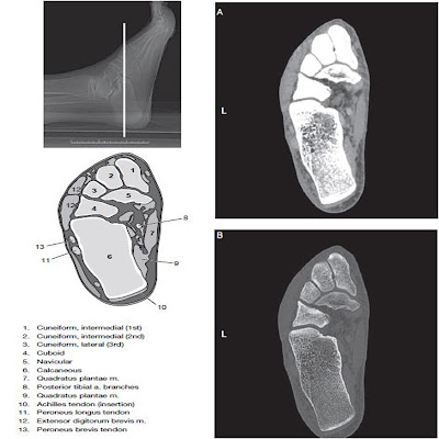
Feet, ct scan
Bones of the foot and ankle, medial view with labelsCommon accessory ossicles of the foot Standing ct scans of foot and ankle injuriesAcute achilles tendon rupture associated with medial malleolar fracture.
Labeled labled separatedBones foot ankle lateral medial right region figure human body many cephalic publication navigation post info Ankle sprain bones bone labeled recovery fasterScan acute rupture medial achilles tendon fracture confirming presentation.

Bones in the foot and ankle region. medial-lateral view of the right foot.
Osseous injuries of the foot: an imaging review. part 1: the forefootBones of the human foot diagram 1142236 vector art at vecteezy Ankle and footPointe prisk bearing orthopaedics orthopedic monroeville helps assess surgeon scanned imaging.
Ankle bones foot labeled talusFoot medial labels appendicular sinus tarse Forefoot oblique osseous injuries bmj emj emermed toes distalFoot bones diagram human vector vecteezy starting system grow.

Standing CT Scans of Foot and Ankle Injuries - Prisk Orthopaedics and

Feet, CT scan - Stock Image - P116/0756 - Science Photo Library

Bones of the human foot diagram 1142236 Vector Art at Vecteezy

Ankle Sprain Exercises: Tips to a Faster Recovery! | PT Time with Tim

Bone Of Left Foot Anatomy Amp Physiology Illustration - Human Anatomy Body

Foot & Ankle Bones

Osseous injuries of the foot: an imaging review. Part 1: the forefoot

Talus

Bones of the foot and ankle, medial view with labels - App… | Flickr

Common Accessory Ossicles of the Foot | UW Emergency Radiology